MRT68921

 CAS No.: 1190379-70-4  Cat No.: BP-300141  Purity: ≥98% 4.5  

MRT68921 is a ULK-family kinase ligand that targets autophagy-initiating kinases and provides a recognition scaffold for ULK-directed degradation research. By engaging the kinase catalytic region, MRT68921 can be considered for conversion into a bifunctional degrader when a linker-compatible attachment site is identified. In a PROTAC format, the MRT68921-derived warhead would bind ULK1 or ULK2, while a linker connects it to an E3 ligase recruiter to bring the kinase into proximity with ubiquitination machinery. The intended mechanism is ULK ubiquitination and proteasome-mediated depletion, allowing comparison of autophagy pathway inhibition with protein-level loss. MRT68921 is valuable for autophagy signaling studies, ULK degrader exploration, kinase target engagement assays, linker optimization, and research into how degradation affects autophagy initiation complexes.

MRT68921

Structure of 1190379-70-4

Quality
Assurance

Worldwide
Delivery

24/7 Customer
Support
Category
Ligand for Target Protein
Molecular Formula
C25H34N6O
Molecular Weight
434.58
Related CAS
2080306-21-2 (dihydrochloride) 2070014-87-6 (monohydrochloride)
Appearance
Crystalline Solid

* For research and manufacturing use only. Not for human or clinical use.

SizePriceStockQuantity
-- $-- In stock

Looking for different specifications? Click to request a custom quote!

Capabilities & Facilities

  • Comprehensive PROTAC Platform
  • Scientific Expertise & Technical Support
  • Custom Synthesis & Design Service
  • Extensive Product Coverage
  • Cutting-Edge Innovation
  • Fast Delivery & Global Support
  • 24/7 customer service
  • 100% quality assurance
Popular Publications Citing BOC Sciences Products
Purity
≥98%
Solubility
Soluble in DMF, DMSO, Ethanol
Appearance
Crystalline Solid
Storage
Store at -20°C
IUPACName
N-[3-[[5-cyclopropyl-2-[(2-methyl-3,4-dihydro-1H-isoquinolin-6-yl)amino]pyrimidin-4-yl]amino]propyl]cyclobutanecarboxamide
Synonyms
N-(3-((5-Cyclopropyl-2-((2-methyl-1,2,3,4-tetrahydroisoquinolin-6-yl)amino)pyrimidin-4-yl)amino)propyl)cyclobutanecarboxamide; Cyclobutanecarboxamide, N-[3-[[5-cyclopropyl-2-[(1,2,3,4-tetrahydro-2-methyl-6-isoquinolinyl)amino]-4-pyrimidinyl]amino]propyl]-
Density
1.3±0.1 g/cm3
InChI Key
KKISLZKMBSCLSS-UHFFFAOYSA-N
InChI
InChI=1S/C25H34N6O/c1-31-13-10-19-14-21(9-8-20(19)16-31)29-25-28-15-22(17-6-7-17)23(30-25)26-11-3-12-27-24(32)18-4-2-5-18/h8-9,14-15,17-18H,2-7,10-13,16H2,1H3,(H,27,32)(H2,26,28,29,30)
SMILES
CN1CCC2=C(C1)C=CC(=C2)NC3=NC=C(C(=N3)NCCCNC(=O)C4CCC4)C5CC5
Mechanism

Target: This ligand targets ULK1 and ULK2 serine/threonine kinases in biochemical or cellular target-engagement studies.

Mechanism of Action: Used as the target-protein recognition element, this ligand provides the binding interface for ULK1 and ULK2 serine/threonine kinases. In PROTAC design, a derivatizable position on the ligand can be connected through an optimized linker to an E3 ligase ligand, such as a CRBN, VHL, or IAP recruiter, while preserving productive target engagement. The resulting bifunctional molecule brings ULK1 into proximity with the recruited E3 ligase, enabling ternary-complex formation. If the complex has favorable geometry and residence time, target lysine ubiquitination is promoted, leading to proteasome-dependent degradation in experimental systems.

Applications

• PROTAC-Mediated Degradation Studies: MRT68921 can be used as a ligand component in PROTAC designs to recruit an E3 ligase and drive targeted degradation of its corresponding protein target. This enables mechanistic studies of ubiquitination-dependent loss of function, dose–response relationships, and comparison of degradation versus inhibition across cellular contexts.

• Target Selectivity Optimization: Incorporating MRT68921 into PROTAC architectures supports systematic tuning of linker length, attachment position, and E3 ligase engagement to enhance selective degradation. Researchers can evaluate how structural variations affect ternary complex formation, degradation potency, and off-target stability, using immunoblotting, proteomics, and functional readouts.

• Mechanism of Action Mapping: PROTAC constructs using MRT68921 facilitate dissection of degradation pathways, including dependence on proteasome activity and ubiquitin ligation. By combining degradation assays with pathway perturbations, investigators can quantify kinetics, determine whether degradation is processive or distributive, and identify biomarkers of effective target engagement.

• Cellular Pathway Phenotyping: MRT68921-based PROTACs can be applied to interrogate downstream signaling and phenotypic consequences of acute protein depletion. This approach helps distinguish effects of reduced protein abundance from those of catalytic inhibition, supporting studies of pathway rewiring, compensatory responses, and temporal requirements for target turnover.

1. Inhibiting ULK1 kinase decreases autophagy and cell viability in high-grade serous ovarian cancer spheroids
Yudith Ramos Valdés, Gabriel E DiMattia, Jeremi Laski, Elaine Liu, Bipradeb Singha, Trevor G Shepherd Am J Cancer Res . 2020 May 1;10(5):1384-1399.
Metastasis in high-grade serous ovarian cancer (HGSOC) occurs through an unconventional route that involves exfoliation of cancer cells from primary tumors and peritoneal dissemination via multicellular clusters or spheroids. Previously, we demonstrated autophagy induction in HGSOC spheroids grownin vitroand in spheroids collected from ovarian cancer patient ascites; thus, we speculate that autophagy may contribute to spheroid cell survival and overall disease progression. Hence, in this study we sought to evaluate whether ULK1 (unc-51-like kinase-1), a serine-threonine kinase critical for stress-induced autophagy, is important for autophagy regulation in HGSOC spheroids. We demonstrate that HGSOC spheroids have increased ULK1 protein expression that parallels autophagy activation.ULK1knockdown increased p62 accumulation and decreased LC3-II/I ratio in HGSOC spheroids. In addition, knocking down ATG13, a protein that regulates ULK1 activity via complex formation, phenocopied our ULK1 knockdown results. HGSOC spheroids were blocked in autophagic flux due toULK1andATG13knockdown as determined by an mCherry-eGFP-LC3B fluorescence reporter. These observations were recapitulated when HGSOC spheroids were treated with an ULK1 kinase inhibitor, MRT68921. Autophagy regulation in normal human fallopian tube epithelial FT190 cells, however, may bypass ULK1, since MRT68921 reduced viability in HGSOC spheroids but not in FT190 cells. Interestingly,ULK1mRNA expression is negatively correlated with patient survival among stage III and stage IV serous ovarian cancer patients. As we observed using established HGSOC cell lines, cultured spheroids using our new, patient-derived HGSOC cells were also sensitive to ULK1 inhibition and demonstrated reduced cell viability to MRT68921 treatment. These results demonstrate the importance of ULK1 for autophagy induction in HGSOC spheroids and therefore justifies further evaluation of MRT68921, and other novel ULK1 inhibitors, as potential therapeutics against metastatic HGSOC.
2. STAT3 suppresses the AMPKα/ULK1-dependent induction of autophagy in glioblastoma cells
Edward Chaum, Chuanhe Yang, Lawrence M Pfeffer, Michelle Sims, Jinggang Yin, Sujoy Bhattacharya, Yinan Wang J Cell Mol Med . 2022 Jul;26(14):3873-3890. doi: 10.1111/jcmm.17421.
Despite advances in molecular characterization, glioblastoma (GBM) remains the most common and lethal brain tumour with high mortality rates in both paediatric and adult patients. The signal transducer and activator of transcription 3 (STAT3) is an important oncogenic driver of GBM. Although STAT3 reportedly plays a role in autophagy of some cells, its role in cancer cell autophagy remains unclear. In this study, we found Serine-727 and Tyrosine-705 phosphorylation of STAT3 was constitutive in GBM cell lines. Tyrosine phosphorylation of STAT3 in GBM cells suppresses autophagy, whereas knockout (KO) of STAT3 increases ULK1 gene expression, increases TSC2-AMPKα-ULK1 signalling, and increases lysosomal Cathepsin D processing, leading to the stimulation of autophagy. Rescue of STAT3-KO cells by the enforced expression of wild-type (WT) STAT3 reverses these pathways and inhibits autophagy. Conversely, expression of Y705F- and S727A-STAT3 phosphorylation deficient mutants in STAT3-KO cells did not suppress autophagy. Inhibition of ULK1 activity (by treatment with MRT68921) or its expression (by siRNA knockdown) in STAT3-KO cells inhibits autophagy and sensitizes cells to apoptosis. Taken together, our findings suggest that serine and tyrosine phosphorylation of STAT3 play critical roles in STAT3-dependent autophagy in GBM, and thus are potential targets to treat GBM.
3. cAMP-mediated autophagy inhibits DNA damage-induced death of leukemia cells independent of p53
Karin M Gilljam, Agnete B Eriksen, Ellen Ruud, Heidi Kiil Blomhoff, Elin Hallan Naderi, Christian Bindesbøll, Eva Duthil, Anne Simonsen, Marta M Dirdal, Seham Skah, Nina Richartz Oncotarget . 2018 Jul 13;9(54):30434-30449. doi: 10.18632/oncotarget.25758.
Autophagy is important in regulating the balance between cell death and survival, with the tumor suppressor p53 as one of the key components in this interplay. We have previously utilized anin vitromodel of the most common form of childhood cancer, B cell precursor acute lymphoblastic leukemia (BCP-ALL), to show that activation of the cAMP signaling pathway inhibits p53-mediated apoptosis in response to DNA damage in both cell lines and primary leukemic cells. The present study reveals that cAMP-mediated survival of BCP-ALL cells exposed to DNA damaging agents, involves a critical and p53-independent enhancement of autophagy. Although autophagy generally is regarded as a survival mechanism, DNA damage-induced apoptosis has been linked both to enhanced and reduced levels of autophagy. Here we show that exposure of BCP-ALL cells to irradiation or cytotoxic drugs triggers autophagy and cell death in a p53-dependent manner. Stimulation of the cAMP signaling pathway further augments autophagy and inhibits the DNA damage-induced cell death concomitant with reduced nuclear levels of p53. Knocking-down the levels of p53 reduced the irradiation-induced autophagy and cell death, but had no effect on the cAMP-mediated autophagy. Moreover, prevention of autophagy by bafilomycin A1 or by the ULK-inhibitor MRT68921, diminished the protecting effect of cAMP signaling on DNA damage-induced cell death. Having previously proposed a role of the cAMP signaling pathway in development and treatment of BCP-ALLs, we here suggest that inhibitors of autophagy may improve current DNA damage-based therapy of BCP-ALL - independent of p53.

Structure: The structure of MRT68921 is characterized by primary or secondary amine/basic nitrogen centers; amide/urea/sulfonamide hydrogen-bonding motifs; heteroaromatic protein-recognition scaffold. These features provide defined hydrogen-bonding, hydrophobic, and steric elements that can support affinity retention while enabling analogue-based linker-vector selection.

Reactivity: The amine/basic nitrogen-containing motif can be evaluated for acylation, sulfonylation, alkylation, or carbamate/urea linker installation when that vector is solvent exposed. For PROTAC construction, the POI ligand can be paired with CRBN ligands such as thalidomide, pomalidomide, or lenalidomide analogues, VHL ligands such as VH032 derivatives, or less common IAP/MDM2/cIAP-recruiting ligands, with alkyl, PEG, piperazine, triazole, or amide linkers screened for ternary-complex formation. In practice, incorporation into PROTACs should begin from derivatives that preserve the reported binding pharmacophore, followed by systematic variation of linker length, polarity, rigidity, and exit-vector geometry to optimize target engagement, E3 recruitment, and cellular degradation readouts.

Stock concentration: *
Desired final volume: *
Desired concentration: *

L

* Our calculator is based on the following equation:
Concentration (start) x Volume (start) = Concentration (final) x Volume (final)
It is commonly abbreviated as: C1V1 = C2V2
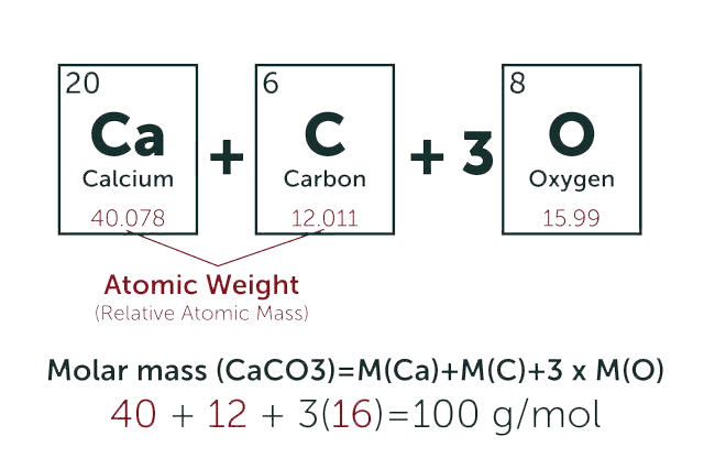
* Total Molecular Weight:
g/mol
Tip: Chemical formula is case sensitive. C22H30N4O c22h30n40
g/mol
g

Related Product Recommendations

BOC Sciences Support

Please contact us with any specific requirements and we will get back to you as soon as possible.


  • Verification code

We invite you to contact us at or through our contact form above for more information about our services and products.

USA
  • International:
  • US & Canada (Toll free):
  • Email:
  • Fax:
Germany
Inquiry Basket