PHA665752

 CAS No.: 477575-56-7  Cat No.: BP-300125  Purity: >98% 4.5  

PHA665752 is a c-Met kinase ligand that binds the catalytic region of the MET receptor tyrosine kinase and provides a selective recognition scaffold for MET-targeted degrader research. In a PROTAC design, the PHA665752-derived moiety would bind c-Met, while a linker connects it to an E3 ligase recruiter to position the receptor kinase near ubiquitination machinery. Productive ternary complex formation is expected to drive c-Met ubiquitination and proteasome-dependent depletion. This approach can help compare receptor kinase inhibition with protein removal and support studies of MET-dependent signaling, receptor scaffold roles, pathway adaptation, and target persistence. PHA665752 is valuable for c-Met degrader exploration, receptor tyrosine kinase biology, linker-exit-vector analysis, target engagement assays, and optimization of degradation selectivity.

PHA665752

Structure of 477575-56-7

Quality
Assurance

Worldwide
Delivery

24/7 Customer
Support
Category
Ligand for Target Protein
Molecular Formula
C32H34Cl2N4O4S
Molecular Weight
641.608
Appearance
light yellow to yellow solid

* For research and manufacturing use only. Not for human or clinical use.

SizePriceStockQuantity
-- $-- In stock

Looking for different specifications? Click to request a custom quote!

Capabilities & Facilities

  • Comprehensive PROTAC Platform
  • Scientific Expertise & Technical Support
  • Custom Synthesis & Design Service
  • Extensive Product Coverage
  • Cutting-Edge Innovation
  • Fast Delivery & Global Support
  • 24/7 customer service
  • 100% quality assurance
Popular Publications Citing BOC Sciences Products
Purity
>98%
Appearance
light yellow to yellow solid
IUPACName
(3Z)-5-[(2,6-dichlorophenyl)methylsulfonyl]-3-[[3,5-dimethyl-4-[(2R)-2-(pyrrolidin-1-ylmethyl)pyrrolidine-1-carbonyl]-1H-pyrrol-2-yl]methylidene]-1H-indol-2-one
Synonyms
PHA-665752; PHA 665752
InChI Key
OYONTEXKYJZFHA-SSHUPFPWSA-N
InChI
InChI=1S/C32H34Cl2N4O4S/c1-19-29(35-20(2)30(19)32(40)38-14-6-7-21(38)17-37-12-3-4-13-37)16-24-23-15-22(10-11-28(23)36-31(24)39)43(41,42)18-25-26(33)8-5-9-27(25)34/h5,8-11,15-16,21,35H,3-4,6-7,12-14,17-18H2,1-2H3,(H,36,39)/b24-16-/t21-/m1/s1
SMILES
CC1=C(NC(=C1C(=O)N2CCCC2CN3CCCC3)C)C=C4C5=C(C=CC(=C5)S(=O)(=O)CC6=C(C=CC=C6Cl)Cl)NC4=O
Mechanism

Target: This ligand targets MET receptor tyrosine kinase in biochemical or cellular target-engagement studies.

Mechanism of Action: Used as the target-protein recognition element, this ligand provides the binding interface for MET receptor tyrosine kinase. In PROTAC design, a derivatizable position on the ligand can be connected through an optimized linker to an E3 ligase ligand, such as a CRBN, VHL, or IAP recruiter, while preserving productive target engagement. The resulting bifunctional molecule brings MET receptor tyrosine kinase into proximity with the recruited E3 ligase, enabling ternary-complex formation. If the complex has favorable geometry and residence time, target lysine ubiquitination is promoted, leading to proteasome-dependent degradation in experimental systems.

Applications

• E3 Ligase Engagement Studies: PHA665752 can be used as a PROTAC ligand to recruit a target protein and drive proximity to an E3 ligase, enabling ubiquitination and degradation. Researchers can evaluate degradation efficiency across cell lines by varying linker length and attachment sites, and quantify knockdown kinetics using immunoblotting or proteomics.

• Targeted Degradation Optimization: Incorporating PHA665752 into PROTAC designs supports systematic optimization of ternary complex formation and cellular potency. Experimental workflows may include assessing dose–response degradation profiles, measuring selectivity against related kinases, and correlating degradation potency with binding affinity and residence time to refine PROTAC architecture.

• Mechanistic Ubiquitination Mapping: PROTACs built with PHA665752 can be applied to dissect ubiquitination mechanisms. By using ubiquitin pathway perturbations (e.g., proteasome inhibition or E1/E3 modulation) and time-resolved assays, investigators can determine whether degradation proceeds through canonical ubiquitin–proteasome routes and identify rate-limiting steps.

• Proteome-Wide Selectivity Profiling: PHA665752-based PROTACs are suitable for exploring off-target degradation and pathway rewiring. Researchers can perform global proteomics after PROTAC treatment to map degradation landscapes, distinguish direct target loss from secondary effects, and establish degradation specificity relative to conventional inhibitors.

• Resistance and Reversibility Analysis: PROTACs using PHA665752 can help study how cells adapt to targeted degradation. Longitudinal experiments can monitor changes in target abundance, E3 ligase expression, and compensatory signaling, while washout and re-treatment designs can evaluate reversibility and the durability of degradation-driven phenotypes.

1.Hepatocyte Growth Factor Receptor c-Met Instructs T Cell Cardiotropism and Promotes T Cell Migration to the Heart via Autocrine Chemokine Release.
Komarowska I1, Coe D1, Wang G1, Haas R1, Mauro C1, Kishore M1, Cooper D1, Nadkarni S1, Fu H1, Steinbruchel DA2, Pitzalis C1, Anderson G3, Bucy P4, Lombardi G5, Breckenridge R6, Marelli-Berg FM7. Immunity. 2015 Jun 16;42(6):1087-99. doi: 10.1016/j.immuni.2015.05.014. Epub 2015 Jun 9.
Effector-T-cell-mediated immunity depends on the efficient localization of antigen-primed lymphocytes to antigen-rich non-lymphoid tissue, which is facilitated by the expression of a unique set of "homing" receptors acquired by memory T cells. We report that engagement of the hepatocyte growth factor (HGF) receptor c-Met by heart-produced HGF during priming in the lymph nodes instructs T cell cardiotropism, which was associated with a specialized homing "signature" (c-Met(+)CCR4(+)CXCR3(+)). c-Met signals facilitated T cell recruitment to the heart via the chemokine receptor CCR5 by inducing autocrine CCR5 ligand release. c-Met triggering was sufficient to support cardiotropic T cell recirculation, while CCR4 and CXCR3 sustained recruitment during heart inflammation. Transient pharmacological blockade of c-Met during T cell priming led to enhanced survival of heart, but not skin, allografts associated with impaired localization of alloreactive T cells to heart grafts.
2.Macrophages programmed by apoptotic cells inhibit epithelial-mesenchymal transition in lung alveolar epithelial cells via PGE2, PGD2, and HGF.
Yoon YS1,2, Lee YJ1,2, Choi YH1,2, Park YM3, Kang JL1,2. Sci Rep. 2016 Feb 15;6:20992. doi: 10.1038/srep20992.
Apoptotic cell clearance results in the release of growth factors and the action of signaling molecules involved in tissue homeostasis maintenance. Here, we investigated whether and how macrophages programmed by apoptotic cells inhibit the TGF-β1-induced Epithelial-mesenchymal transition (EMT) process in lung alveolar epithelial cells. Treatment with conditioned medium derived from macrophages exposed to apoptotic cells, but not viable or necrotic cells, inhibited TGF-β1-induced EMT, including loss of E-cadherin, synthesis of N-cadherin and α-smooth muscle actin, and induction of EMT-activating transcription factors, such as Snail1/2, Zeb1/2, and Twist1. Exposure of macrophages to cyclooxygenase (COX-2) inhibitors (NS-398 and COX-2 siRNA) or RhoA/Rho kinase inhibitors (Y-27632 and RhoA siRNA) and LA-4 cells to antagonists of prostaglandin E2 (PGE2) receptor (EP4 [AH-23848]), PGD2 receptors (DP1 [BW-A868C] and DP2 [BAY-u3405]), or the hepatocyte growth factor (HGF) receptor c-Met (PHA-665752), reversed EMT inhibition by the conditioned medium.
3.Hepatic Radiofrequency Ablation-induced Stimulation of Distant Tumor Growth Is Suppressed by c-Met Inhibition.
Ahmed M1, Kumar G1, Moussa M1, Wang Y1, Rozenblum N1, Galun E1, Goldberg SN1. Radiology. 2016 Apr;279(1):103-17. doi: 10.1148/radiol.2015150080. Epub 2015 Sep 29.
Purpose To elucidate how hepatic radiofrequency (RF) ablation affects distant extrahepatic tumor growth by means of two key molecular pathways. Materials and Methods Rats were used in this institutional animal care and use committee-approved study. First, the effect of hepatic RF ablation on distant subcutaneous in situ R3230 and MATBIII breast tumors was evaluated. Animals were randomly assigned to standardized RF ablation, sham procedure, or no treatment. Tumor growth rate was measured for 3½ to 7 days. Then, tissue was harvested for Ki-67 proliferative indexes and CD34 microvascular density. Second, hepatic RF ablation was performed for hepatocyte growth factor (HGF), vascular endothelial growth factor (VEGF), and c-Met receptor expression measurement in periablational rim, serum, and distant tumor 24 hours to 7 days after ablation. Third, hepatic RF ablation was combined with either a c-Met inhibitor (PHA-665752) or VEGF receptor inhibitor (semaxanib) and compared with sham or drug alone arms to assess distant tumor growth and growth factor levels.
4.The EGFR/ErbB3 Pathway Acts as a Compensatory Survival Mechanism upon c-Met Inhibition in Human c-Met+ Hepatocellular Carcinoma.
Steinway SN1, Dang H1, You H1, Rountree CB1, Ding W1. PLoS One. 2015 May 22;10(5):e0128159. doi: 10.1371/journal.pone.0128159. eCollection 2015.
BACKGROUND: c-Met, a high-affinity receptor for Hepatocyte Growth Factor (HGF), plays a critical role in tumor growth, invasion, and metastasis. Hepatocellular carcinoma (HCC) patients with activated HGF/c-Met signaling have a significantly worse prognosis. Targeted therapies using c-Met tyrosine kinase inhibitors are currently in clinical trials for HCC, although receptor tyrosine kinase inhibition in other cancers has demonstrated early success. Unfortunately, therapeutic effect is frequently not durable due to acquired resistance.
ConcentrationVolumeMass1 mg5 mg10 mg
1 mM1.5586 mL7.7929 mL15.5858 mL
5 mM0.3117 mL1.5586 mL3.1172 mL
10 mM0.1559 mL0.7793 mL1.5586 mL
50 mM0.0312 mL0.1559 mL0.3117 mL

PHA665752 is a c-Met kinase ligand scaffold for c-Met-directed degrader design. Linker installation should preserve the heteroaryl kinase-binding core and use a validated solvent-exposed vector.

Structure: PHA665752 is a c-Met kinase ligand scaffold containing a quinoline/quinazoline-like heteroaryl system, halogenated aryl substitution, and an amide-linked heterocyclic side chain. The structure includes multiple aromatic rings, heteroaryl nitrogens, and polar amide functionality.

Reactivity: For c-Met-directed PROTAC construction, the heteroaryl kinase-binding core should be preserved while linker installation is explored from solvent-exposed amide or heterocyclic side-chain vectors. Alkyl, PEG, amide, carbamate, or piperazine-containing linkers may be coupled to CRBN, VHL, or IAP ligands in appropriate analogs. Modification should be benchmarked against c-Met binding because changes near the quinoline/heteroaryl core may disrupt recognition.

Stock concentration: *
Desired final volume: *
Desired concentration: *

L

* Our calculator is based on the following equation:
Concentration (start) x Volume (start) = Concentration (final) x Volume (final)
It is commonly abbreviated as: C1V1 = C2V2
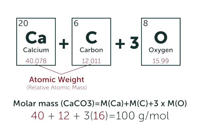
* Total Molecular Weight:
g/mol
Tip: Chemical formula is case sensitive. C22H30N4O c22h30n40
g/mol
g

Related Product Recommendations

BOC Sciences Support

Please contact us with any specific requirements and we will get back to you as soon as possible.


  • Verification code

We invite you to contact us at or through our contact form above for more information about our services and products.

USA
  • International:
  • US & Canada (Toll free):
  • Email:
  • Fax:
Germany
Inquiry Basket